Welcome to the fascinating realm of mitosis in onion root tip lab answers, where we delve into the intricate process of cell division that governs the growth and development of all living organisms. This lab provides a hands-on opportunity to witness the dynamic stages of mitosis and gain a deeper understanding of its significance in plant biology.
As we embark on this exploration, we will unravel the secrets of onion root tips, which serve as an ideal model for studying mitosis due to their rapid cell division rate. Through a series of carefully designed experiments, we will identify and count cells in each stage of mitosis, calculate the mitotic index, and analyze the data to gain insights into the regulation and control of cell division.
1. Mitosis in Onion Root Tip Lab
Background and Introduction

Mitosis is a fundamental process in cell division, essential for the growth and development of all living organisms. It is the process by which a single cell divides into two identical daughter cells, ensuring the accurate transmission of genetic material.
Onion root tips serve as an ideal model for studying mitosis due to their rapid cell division and the presence of numerous mitotic figures. The cells in the root tip are easily accessible and can be stained to visualize the different stages of mitosis.
2. Materials and Methods
Materials:
- Onion bulb with roots
- Hydrochloric acid (HCl)
- Orcein stain
- Microscope slides and coverslips
- Forceps
- Microscope
Procedure:
- Cut off the root tips (1-2 cm) from the onion bulb and place them in a petri dish containing HCl for 15 minutes.
- Rinse the root tips with water and transfer them to a slide.
- Add a drop of orcein stain to the root tips and heat gently for 2-3 minutes.
- Place a coverslip over the root tips and gently press down to spread the cells.
- Observe the stained root tips under a microscope.
3. Observations and Data Collection
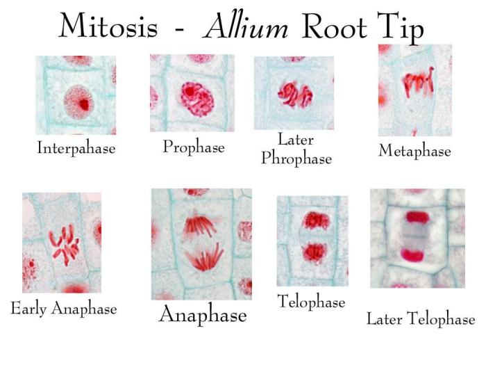
Under the microscope, the different stages of mitosis can be observed:
- Interphase:The cell is not dividing and the chromosomes are not visible.
- Prophase:The chromosomes condense and become visible. The nuclear membrane begins to break down.
- Metaphase:The chromosomes line up at the equator of the cell.
- Anaphase:The chromosomes separate and move to opposite poles of the cell.
- Telophase:Two new nuclear membranes form around the chromosomes. The cell membrane pinches in the middle, dividing the cell into two daughter cells.
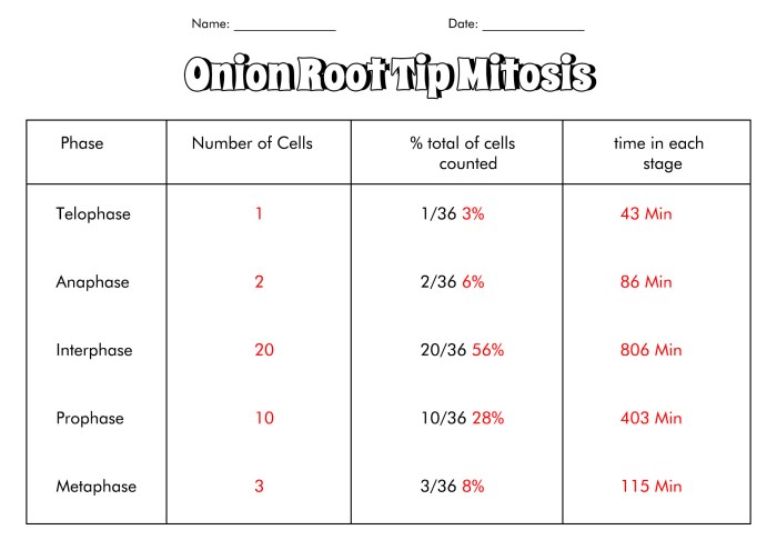
To count the cells in each stage of mitosis, use a tally counter or create a table or spreadsheet with columns for each stage.
4. Data Analysis
Calculate the mitotic index by dividing the number of cells in mitosis by the total number of cells observed and multiplying by 100:
Mitotic index = (Number of cells in mitosis / Total number of cells) x 100
The mitotic index indicates the percentage of cells in the onion root tip sample that are actively dividing.
Create a graph or chart to represent the distribution of cells in different stages of mitosis. This will help visualize the progression of the cell cycle.
5. Discussion and Conclusion, Mitosis in onion root tip lab answers
The mitosis in onion root tip lab provides a valuable opportunity to observe and study the process of cell division. The results of the lab can be used to calculate the mitotic index and to create a graphical representation of the cell cycle.
The mitotic index can provide insights into the growth rate and metabolic activity of the onion root tip. A high mitotic index indicates a rapidly growing tissue, while a low mitotic index indicates a slower growth rate.
The study of mitosis is essential for understanding the growth and development of plants and animals. Mitosis ensures the accurate transmission of genetic material from one generation to the next, and it plays a crucial role in maintaining the health and integrity of organisms.
Popular Questions: Mitosis In Onion Root Tip Lab Answers
What is the significance of studying mitosis in onion root tips?
Onion root tips are an excellent model for studying mitosis because they have a high rate of cell division, making it easier to observe the different stages of mitosis under a microscope.
How can we identify and count cells in each stage of mitosis?
Cells in different stages of mitosis can be identified based on their characteristic chromosomal structures. By counting the number of cells in each stage, we can determine the mitotic index, which provides insights into the rate of cell division.
What is the mitotic index and how is it calculated?
The mitotic index is the percentage of cells in a population that are undergoing mitosis. It is calculated by dividing the number of cells in mitosis by the total number of cells counted.


